

DIGITAL PATHOLOGY
Aperio CS2
Create high-quality digital slides with the ultra-compact Aperio CS2 image capture device from your desktop. With a five-slide capacity and 20X and 40x magnification capabilities, the Aperio CS2 is a highly reliable workhorse for medium-volume sites.
The intuitive interface and easy-to-use design consistently provides rapid creation of high resolution digital slides, with >98% first time scan success rate.
The Aperio CS2 runs on the Microsoft Windows 10 operating system.

Aperio AT2
We understand that your slides are more than just glass, they hold vital information for your work. With its compact size, high capacity autoloader and optimal image quality, the Aperio AT2 slide scanner has set new standards in digital pathology.
Aperio AT2 consistently delivers precise, whole slide images with an unparalleled, low rescan rate. Slides are available for remote viewing in less than a minute. With an easy-to-use Pathologist's cockpit, coupled with easy integration into laboratory information systems (LIS), the Aperio AT2 provides an ideal platform for research institutions.
The Aperio AT2 runs on the Microsoft Windows 10 operating system.

Aperio VERSA
The Aperio VERSA is a comprehensive digital pathology scanner, designed and developed to support the diverse imaging needs of cutting edge research facilities. The combination scanner is optimized for precision scanning of brightfield and fluorescent samples, with the accuracy and resolution required for FISH.
From tissue-based and proteomic markers, to subcellular, molecular and in-situ hybridization probes, the Aperio VERSA delivers high-resolution, reliable imaging. Users can create a permanent record of their research – even faint fluorescent samples, which are often subject to fading.
The Aperio VERSA is ideal for scanning multiplex slides at any magnification from 5x to 63x for the whole slide without the need for spectral imaging.

Aperio GT 450
The Aperio GT 450 enables histotechnicians to complete scanning tasks quickly and with confidence leveraging a 32-second scan speed*. Output 81 slides/hr at 40x* delivering high quality images with Leica optics and with an IT architecture that is secure and scalable. From the pathology lab to the IT room, the Aperio GT 450 is designed to scale up digital pathology operations.
-
Improve case turnaround with no touch during scanning and continous loading of the racks directly from the HistoCore Workstation (Stainer & Coverslipper)
-
Help increase IT security and control with a dedicated SAM (Scanner Admin Manager) server and software that allows you to set up and monitor multiple Aperio GT 450s at a time. No more single workstations for each scanner
-
Ensure excellent image quality with Leica optics

Aperio eSlide Manager
Aperio eSlide Manager provides full scalability and optimal performance from single-site installations to multi-site global hub and spoke networks. With dedicated workflows for research and biopharma coupled with an intuitive interface, Aperio eSlide Manager is the ideal solution to meet the diverse needs of both entry-level and enterprise digital pathology users.

Aperio ImageScope
Join the thousands who use our freely downloadable ImageScope viewing software—experience rapid access to crisp, true-color digital slide images to which you can adjust magnification, pan and zoom, compare different stains, annotate areas of interest, perform image analysis, and more.
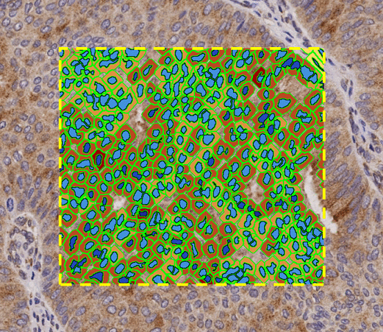
IHC – Brightfield Immunohistochemistry Image Analysis
The Aperio Image Analysis IHC menu provides a broad range of solutions for quantification of single and multiplex tissue staining. Flexible algorithms are easily optimized for diverse brightfield chromogens, enabling you to customize the analysis to match your unique research needs. Accurately identify protein biomarker expression at the tissue, cellular or subcellular level.

Immunofluorescence – Fluorescent Immunohistochemistry Image Analysis
The Aperio Image Analysis Immunofluorescence menu offers solutions for measurement of single and multiplex tissue staining. Quantify expression of fluorescently-labeled protein biomarkers at the tissue, cellular or subcellular level. Algorithms can be customized for a variety of fluorescent dyes, allowing each algorithm to be used for specific, diverse research requirements.

ISH & FISH – Brightfield & Fluorescence In Situ Hybridization Enumeration
Molecular biomarkers can be time-consuming to quantify manually. Aperio Image Analysis offers solutions for automated detection and counting of target signals, with options for brightfield and fluorescence. Algorithms are flexible enough to handle single-plex or multiplex analysis, and can be easily optimized to meet a variety of end-user requirements. Measure RNA or DNA biomarker expression at the tissue, cellular, or subcellular level.

Pattern Recognition – Morphology & Tissue Microarray Analysis Identification Tools
Aperio Image Analysis provides tools to automate tissue identification tasks that would otherwise require time-consuming manual work. Use annotations and templates to flexibly guide automated recognition of histological features or TMA spots, improving identification accuracy and reducing manual intervention.

Clinical Analysis – Validated Algorithms to Support Decision Making
Aperio Image Analysis tools for clinical image analysis, designed to support clinical decisions while reducing inter- and intra-observer variability. Computer-aided image analysis provides a powerful aid to diagnosis, and supports workflows that may be eligible for reimbursement.

Scan – Aperio Digital Pathology Slide Scanners
The digital slide scanning range from Leica Biosystems offers exceptional image quality, speed and reliability for whole slide imaging, making Aperio scanners the optimal choice for healthcare and research professionals.
Patented capture technology and world-leading optics ensures precise high-resolution capture of slide details for brightfield, fluorescent or FISH specimens. Further extend your digital pathology capabilities with interoperable solutions in the Manage and Analyze portfolios.